 The light microscope, also known as a compound microscope or compound light microscope, allows you to see tiny objects and creatures at about 1,000 times their normal size. Students, scientists, researchers and law enforcement agencies use these microscopes in their daily work. Light microscopes are available in a range of prices and qualities from a basic student microscope to a professional model with additional light sources, eyepieces or objective lenses.
Thursday, January 24, 2013
Light Microscope Uses By Kathryn Hulick
The light microscope, also known as a compound microscope or compound light microscope, allows you to see tiny objects and creatures at about 1,000 times their normal size. Students, scientists, researchers and law enforcement agencies use these microscopes in their daily work. Light microscopes are available in a range of prices and qualities from a basic student microscope to a professional model with additional light sources, eyepieces or objective lenses.Infinity Corrected Optics from MicroscopeWorld
| Useful Article about lenses from MicroscopeWorld What are Infinity Corrected Optics? | |
Before Infinity corrected objectives were introduced about a decade ago, all microscopes had a fixed tube length. Microscopes that do not unilitze an infinity corrected optical system have a specified tube length - that is, a set distance from the nosepiece where the objective is attached to the point where the ocular sits in the eyetube. The Royal Microscopical Society standardized microscope tube length at 160mm during the nineteenth century and this standard was accepted for over 100 years.
When optical accessories such as a vertical illuminator or a polarizing accessory are added into the light path of a fixed tube length microscope the once perfectly corrected optical system now has an effective tube length greater than 160mm. In order to adjust for the change in tube length manufacturers were forced to place additional optical elements into the accessories in order to re-establish the 160mm tube length. This usually resulted in increased magnification and reduced light.
German microscope manufacturer Reichert started experimenting with Infinity corrected optical systems in the 1930s and were followed later by Zeiss and Leica. However, the infinity optical system did not become common place until the 1980s.
Infinity corrected optical systems are used in research laboratory microscopes and industrial metallurgical microscopes. These microscope systems have an image distance that is set to infinity. A tube lens is placed within the body tube between the objective and the eyepieces to produce the intermediate image. The Infinity optical system allows auxiliary components (such as illuminators, polarizers, etc.) to be added into the parallel optical path between the objective and the tube lens with only a minimal effect on focus.
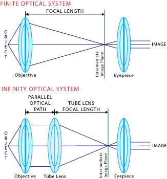
The tube length in infinity corrected optical systems (also known as the focal length) ranges from 160 - 200mm. Meiji, Leica and Nikon all have a focal length of 200mm, Olympus uses 180mm, and Zeiss 165mm
| |
The upside of having Infinity Space in your microscope objective
| |
| The space between the objective and the tube lens (shown in the image above labelled as "Parallel optical path") can be referred to as Infinity Space and provides a path of parallel light where optical components can be placed without the introduction of spherical abberration or modification of the objective's working distance. One benefit in using Infinity Corrected Optics is that parfocality between different objectives can still be maintained, even when one or more auxiliary compenents (such as illuminators or polarizers) are added into the optical path. Another benefit of the Infinity Space is that accessories can be created to produce exactly 1x magnification without altering the alignment between the objective and tube lens. This allows the user to compare specimens using a combination of optical techniques, such as phase contrast. This works because when the optical accessories are placed within that "Infinity Space" they do not shift the location or the focal point of the image. | |
Infinity Optics Defined
| |
| The term "Infinity Optics" actually refers to the production of a flux of parallel light rays after passing through the objective. It would seem that the term refers to the Infinte Space in the microscope for adding additional components, but this is not the true definition. | |
Monday, January 14, 2013
Principles of Microscopy from Stephen M. Wolniak
Stephen M. Wolniak
Professor
Department of Cell Biology & Molecular Genetics
University of Maryland
College Park, Maryland, 20742swolniak@umd.edu

Brightfield microscopy
The microscope that is available to you for general use in this laboratory is a sophisticated optical instrument that can provide you with high-resolution images of a variety of specimens. Image quality is based largely on your ability to use the microscope properly. Below you will find some basic information that you have probably heard before, but information that is rarely presented in a thorough way.
- Resolution -
The magnification of small things is a necessary facet of biological research, but the fine detail in cells and in subcellular components requires that any imaging system be capable of providing spatial information across small distances. Resolution is defined as the ability to distinguish two very small and closely-spaced objects as separate entities. Resolution is best when the distance separating the two tiny objects is small. Resolution is determined by certain physical parameters that include the wavelength of light, and the light-gathering power of the objective and condenser lenses. A simple mathematical equation defines the smallest distance (dmin) separating the two very small objects:
dmin = 1.22 x wavelength / N.A. objective + N.A. condenser
N.A. (Numerical Aperture) is a mathematical calculation of the light-gathering capabilities of a lens. The N.A. of each objective lens is inscribed in the metal tube, and ranges from 0.25-1.4. The higher the N.A., the better the light-gathering properties of the lens, and the better the resolution. Higher N.A. values also mean shorter working distances (you have to get the lens closer to the object). N.A. values above 1.0 also indicate that the lens is used with some immersion fluid, such as immersion oil.
From the equation above, you should be aware that the N.A. of the condenser is as important as the N.A. of the objective lens in determining resolution. It is for this reason that closure of the condenser diaphragm results in a loss of resolution. In practice, at full aperture and with good oil immersion lenses (N.A. 1.4 for both the condenser and the objective) it is possible to be able to resolve slightly better than 0.2 µm. From the equation above, it should also be clear that shorter wavelength light (bluer light) will provide you with better resolution (smaller dmin values). However, there are practical considerations in how short the wavelength can be. In the early 1950's, a UV microscope was designed, but required quartz objectives and a specialized imaging device. The quartz lenses provided slightly better resolution (dmin = 0.1 µm), but image quality suffered from an inability on the part of the manufacturers to correct for aberrations caused by the quartz. The human eye is best adapted for green light and our ability to see detail may be compromised somewhat with the use of blue or violet. Most manufacturers of microscopes correct their simplest lenses (achromats) for green light.
History of the Microscope How the light microscope evolved.
 During that historic period known as the Renaissance, after the "dark" Middle Ages, there occurred the inventions of printing, gunpowder and the mariner's compass, followed by the discovery of America. Equally remarkable was the invention of the light microscope: an instrument that enables the human eye, by means of a lens or combinations of lenses, to observe enlarged images of tiny objects. It made visible the fascinating details of worlds within worlds.
Invention of Glass Lenses
Long before, in the hazy unrecorded past, someone picked up a piece of transparent crystal thicker in the middle than at the edges, looked through it, and discovered that it made things look larger. Someone also found that such a crystal would focus the sun's rays and set fire to a piece of parchment or cloth. Magnifiers and "burning glasses" or "magnifying glasses" are mentioned in the writings of Seneca and Pliny the Elder, Roman philosophers during the first century A. D., but apparently they were not used much until the invention of spectacles, toward the end of the 13th century. They were named lenses because they are shaped like the seeds of a lentil.
The earliest simple microscope was merely a tube with a plate for the object at one end and, at the other, a lens which gave a magnification less than ten diameters -- ten times the actual size. These excited general wonder when used to view fleas or tiny creeping things and so were dubbed "flea glasses."
Birth of the Light Microscope
About 1590, two Dutch spectacle makers, Zaccharias Janssen and his son Hans, while experimenting with several lenses in a tube, discovered that nearby objects appeared greatly enlarged. That was the forerunner of the compound microscope and of the telescope. In 1609,Galileo, father of modern physics and astronomy, heard of these early experiments, worked out the principles of lenses, and made a much better instrument with a focusing device.Anton van Leeuwenhoek (1632-1723)
The father of microscopy, Anton van Leeuwenhoek of Holland, started as an apprentice in a dry goods store where magnifying glasses were used to count the threads in cloth. He taught himself new methods for grinding and polishing tiny lenses of great curvature which gave magnifications up to 270 diameters, the finest known at that time. These led to the building of his microscopes and the biological discoveries for which he is famous. He was the first to see and describe bacteria, yeast plants, the teeming life in a drop of water, and the circulation of blood corpuscles in capillaries. During a long life he used his lenses to make pioneer studies on an extraordinary variety of things, both living and non living, and reported his findings in over a hundred letters to the Royal Society of England and the French Academy.Robert Hooke
Robert Hooke, the English father of microscopy, re-confirmed Anton van Leeuwenhoek's discoveries of the existence of tiny living organisms in a drop of water. Hooke made a copy of Leeuwenhoek's light microscope and then improved upon his design.Charles A. Spencer
Later, few major improvements were made until the middle of the 19th century. Then several European countries began to manufacture fine optical equipment but none finer than the marvelous instruments built by the American, Charles A. Spencer, and the industry he founded. Present day instruments, changed but little, give magnifications up to 1250 diameters with ordinary light and up to 5000 with blue light.Beyond the Light Microscope
A light microscope, even one with perfect lenses and perfect illumination, simply cannot be used to distinguish objects that are smaller than half the wavelength of light. White light has an average wavelength of 0.55 micrometers, half of which is 0.275 micrometers. (One micrometer is a thousandth of a millimeter, and there are about 25,000 micrometers to an inch. Micrometers are also called microns.) Any two lines that are closer together than 0.275 micrometers will be seen as a single line, and any object with a diameter smaller than 0.275 micrometers will be invisible or, at best, show up as a blur. To see tiny particles under a microscope, scientists must bypass light altogether and use a different sort of "illumination," one with a shorter wavelength.Useful Microscope Magnification Information from MicroscopeWorld
| Microscope Magnification | ||
A microscope's total magnification is a combination of the eyepieces and the objective lens. For example, a biological microscope with 10x eyepieces and a 40x objective has 400x magnification. There are however, a few limits to the amount of total magnification that can be reached before empty magnification comes into play. Empty magnification occurs when the image continues to be enlarged, but no additional detail is resolved. This is often the case when higher magnification eyepieces are used. In order to avoid empty magnification, there are a few simple steps that are helpful to follow.
| ||
Eyepiece and Objective Combinations for Optimal Magnification
| ||
When selecting a combination of eyepieces and objective lenses for the optimal magnification, without ending up with "empty magnification" it is important to consider the numerical aperature (NA) of the objective. The numerical aperture of a microscope objective defines the objective's resolution. Each microscope objective has a minimum and maximum magnification necessary for the details in an image to be resolved. A simple formula for the minimum value is (500 x NA). And for the maximum magnification (1000 x NA). Magnifications higher than this value will result in empty magnification, or an image that has a poor resolution. The table below shows some typical NA values with their corresponding objective and provides a range of useful magnification combinations. The blank boxes in the table would provide empty magnification and should be avoided. For example, pairing 20x eyepieces with a 100x objective would not provide good resolution and would result in empty magnification. To determine this, we took 1.25NA x 1000 = 1250 magnification maximum. However, the combination of the 100x objective x 20x eyepieces = 2000, which is above the maximum magnification.
Range of Useful Magnification based on NA of Objectives
 | ||
Thursday, January 3, 2013
Meiji Stereo Microscope Review by Alan Plante:

Reviewed by Alan Plante
The Meiji Techno brand stereo microscopes are becoming increasingly popular among rockhounds who study and/or photograph micro-crystals. Meiji 'scopes are available in either binocular or trinocular models. The binocular models are the ones of choice for people not interested in taking photographs of their micros; while the trinocular - or "third tube" - models are popular among people who want to attach a camera and take pictures through the 'scope.
The most popular models are the EMZ-5, EMZ-5TR and EMZ-8TR, the first of which is a binocular 'scope, the other two being trinocular, for use with cameras. Shown above are two views of the EMZ-8TR. Meiji also makes a model called the RZ-TR, a "fully dedicated" photographic microscope which has a trinocular head and a built in adjustable iris for controlling depth-of-field in photographs.
The basic EMZ5 unit is a binocular stereo zoom 'scope with stand which comes with an 0.7X to 4.5X objective lens (the lower lens) and 10X eyepieces, giving it a magnification range of 9X to 45X. There are also 12.5X, 15X, 20X or 30X eyepieces available for this model. A 15X set gives a magnification range of 10.5X to 67.5X; the 20X eyepieces give a range of 14X to 90X; and the 30X set gives a range of 21X to 135.0X. The optics are "wide field" types, and are of high quality, giving a wide, crisp, and bright image with very good inherent depth-of-field - the portion of the image seen which is in focus from front-to back, or - simply - how much of the image can be brought into focus at one time. A basic 'scope and stand with 10X eyepieces costs around $1,400.00 The 12.5X, 15X and 20X eyepieces are about $140.00 per set (same as the 10X) and the 30X set runs about $160.00. (Prices may vary - those provided are approximate and as of Fall 2001.) These oculars have an inherent "high eyepoint"; and there is also a special 15X model available with a particularly high eyepoint.
Subscribe to:
Comments (Atom)